An Overview of Cervical Degenerative Disc Disease
By reading the anatomy section of this web site, you can understand how the neck is constructed. The discs are wedged in between the vertebral bodies in the front of the spine and the paired facets join the two vertebra in the back of the spine. These movable areas can wear out and cause neck pain. The neck pain from the wear of these structures may be located in the center of the neck, the side of the neck and also may radiate into the top or middle of the shoulders. If the pain radiates down the arm or up the back of the head, this is not disc or facet pain but nerve pain and can occur in conjunction with neck pain. It results from compression of the nerve root which is a byproduct of cervical degenerative disc disease and cervical degenerative facet disease. See the section on nerve root compression for more information on this condition.
The causes of neck pain can often be linked to degenerative changes within the cervical spine. To explain cervical degenerative disc disease and cervical degenerative facet disease, it’s important to understand first, what is a degenerative disc?
Mechanically, everything in the cervical spine revolves around the disc. The disc functions like a shock absorber. This structure absorbs impact and still allows for motion of the spine. It also has to have restraints to prevent damage to itself and the other spinal structures such as the nerves and facets. These requirements create some major demands. In appearance, the disc looks like a jelly donut. The jelly (called the nucleus) is made of sugars attached to a protein backbone called a proteoglycan. This structure allows it to act like a giant sponge. The jelly pulls in water from the body of the vertebra to create a high-pressure interior matrix (think of the jelly as the air pressure in a tire).
The outside of the donut is made up of about twenty to thirty rings of collagen, called the annulus, just like the plies of a tire. These rings are normally quite tough. Each layer of these rings alternates in angulation in their attachment to the bone of the vertebra.
The endplates of the disc separate the bone of the vertebral body from the interior of the disc. They are made of hyaline cartilage-the same cartilage that lines the hip and knee joints. This material creates a barrier to nutrients and oxygen entering and exiting the disc.
An additional anatomic point that the cervical spine has but the thoracic and lumbar spines don’t is the uncovertebral joint. As noted in the anatomy section, this joint is not present in the infant and slowly develops with maturity. The reason this joint develops lies with the design of the surfaces of the vertebral body.
Instead of having flat parallel surfaces that the disc attaches to (like the thoracic and lumbar vertebra), the sides of the body of the cervical vertebra are sloped upwards like a smile when viewed from the front. The disc is wedged in between the two vertebral bodies. Over time, the two surfaces of the vertebral bodies touch. Since they are lined with cartilage, they form an articulation-essentially a new joint.
Blood Supply and Imbibition
Cervical degenerative disc disease and cervical degenerative facet disease have problems that exist with the design of the disc that causes the “disease” we know today as degenerative disc disease or DDD. The first problem is that the blood supply, for all intensive purposes, disappears from the disc by about the age of eight. This means that collectively, the discs are the largest structures in the body that have no blood supply.
Without a blood supply, there is very poor oxygen penetration into the interior of the disc. The only fluids that can be exchanged are under hydrostatic and osmotic pressure. This means that motion of the disc exchanges fluids similarly to a squeezing a sponge under water and releasing it. The water and nutrients that this material absorbs and releases, transfers through the endplate of the vertebra under a process called imbibition.
The fluids that are transferred into the disc are poor in oxygen and limited in nutrients. This creates a problem for the living cells inside the disc. These cells produce the glycoproteins, which make up the nucleus, (the gel or sponge) and they need oxygen to function well. Without oxygen, these cells become much less effective keeping up with maintaining the jelly. Without the production and maintenance of these gel proteins, the pressure in the disc drops with age.
Pressure Drop
When spine surgeons are asked, “what is a degenerative disc?”, it helps to use the following analogy. When the pressure drops inside the disc, this is similar to letting some air out of a car tire. As the pressure in the car tire drops, the sidewalls bulge outward from the weight of the car. When you then try to drive on a partially filled tire, there is less stability to hold the road, especially with unpredicted maneuvers such as a quick turn to avoid an obstacle or driving over an unexpected speed bump. Just as the car has trouble holding the road, a degenerative disc is much less resistant to abnormal movements and these motions can tear the disc wall. An injury or unexpected lift or fall may injure the disc. When the disc loses height, the two vertebral bodies approximate or come closer together. The uncovertebral joint sitting on each side of the disc space undergoes increased pressure and simply wears out.
The uncovertebral joint-like any other joint will develop bone spurs with this undue pressure. Since this joint makes up the front wall of the foramen or hole that the nerve exits, this bone spur can crowd out the nerve root that sits immediately behind the spur. If the nerve root becomes pinched – it will swell which obviously compounds the problem. A swollen root won’t fit well into an already narrowed exit hole. This drop in pressure within the cervical spine is one of the most common causes of neck pain in adults.
Annular Tears and Nociceptors
If a tear occurs, without a blood supply the disc can’t heal if injured. All injuries to the disc therefore are cumulative or add up. To say it another way, any injury to the disc is essentially, a permanent injury. A tear of the collagen in the donut won’t repair itself. The cells lining the outside wall of the annulus attempt to repair the outer defect but are unsuccessful and actually get in the way.
There is a tenuous blood supply in the outer 1/3 of the disc. Tears that go out to here will attempt to heal but the scar tissue laid down is not nearly as strong as the collagen fibers it attempts to replace. To make matters worse, any blood vessels that grow into the torn fibers of the disc carry along with them new pain fibers. These fibers are highly sensitive and another major cause of pain sensitization of the disc.
The back portion of the disc (posterior annulus) is full of pain sensors called nocicepters. When a tear occurs in the annulus of the disc, these pain receptors come in contact with the nucleus of the disc. The pain receptors themselves then become inflamed and much more easily transmit pain signals. This is a common cause of neck pain.
Your Mom and Dad
The second problem with cervical degenerative disc disease and cervical degenerative facet disease is how you picked your mom and dad— genetics. This has to do with the type of collagen that makes up the plies of the tire (the annulus.) The collagen in the annulus is not uniform. Some fibers are pliable and resilient and can undergo multiple loading cycles without any wear just like thick bungee cords. Other types of collagen are unfortunately brittle, like wire coat hangers. You can bend these only a limited amount of times before they break from fatigue failure.
Good examples of this genetic relationship are the variety of patients I am asked to see. Some are injured on the ski slopes. Eighty and occasionally ninety years olds (and yes they ski in Vail) will sporadically develop pain in their backs and have an MRI performed through the emergency room. There are times these octo and nonagenarians have perfect appearing discs (they obviously picked the right parents.) On the other end of the spectrum are the unfortunate twelve year olds in my practice with severe cervical degenerative disc disease.
Remember Wiring!
There is a big caveat here. Remember, just because the patient may have cervical degenerative disc disease and degenerative facet disease does not mean they are going to have symptoms of neck pain, arthritis of the neck or other ailments such as a slipped disc. A study out of Emery University noted that more than 70% of individuals without neck or arm pain had degenerative changes in their discs. These people had no symptoms. I am always reminded of this when I see older NFL players with horrible looking spines and minimal to no pain. This leads into the theory of wiring.
There are 2 groups of patients with the same problems found on x-rays and MRI. One group has no symptoms and the other may be incapacitated with the same type of degenerative spine. It appears that some individuals are “wired” to have pain and others are not. This effect has to do with the sensitivity of the central nervous system and the number of pain receptors present and active in the area of injury. Those who were found to have some variation of arthritis of the neck, a slipped disc, facet arthritis, etc. have specific conditions that are the cause of neck pain.
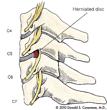
(Click to Enlarge Image) This illustration is a side view of the cervical spine (neck). Here you can see the vertebral bodies with the discs in the front of the spine and the facets in the rear. The nerve roots exit through the sides of the spine.
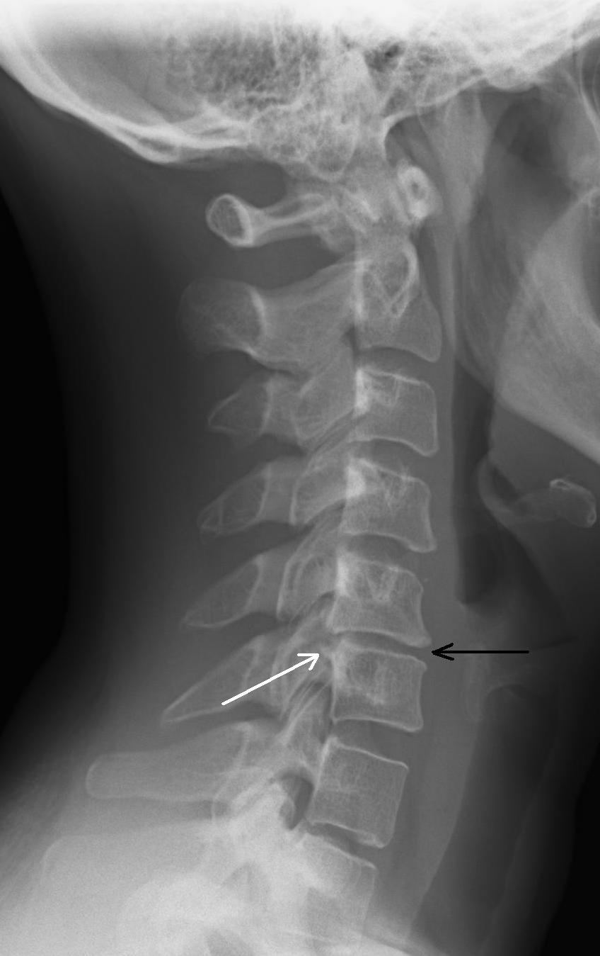
(Click to Enlarge Image) This is a side view of a cervical spine (neck) with a degenerative disc at C5-6. The black arrow points to the narrowed disc and the white arrow points to the bone spur projecting off the back of the disc into the spinal canal.
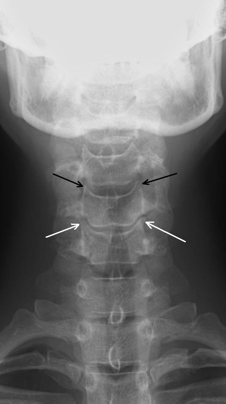
(Click to Enlarge Image) This is a front to back view of a cervical spine (neck) X-ray. The black arrows point to the small pyramid shaped normal uncovertebral joints and the white arrows point to the degenerative uncovertebral joints. Compare the normal to the abnormal to see the enlargement of these joints with a “mushroom head” on one.
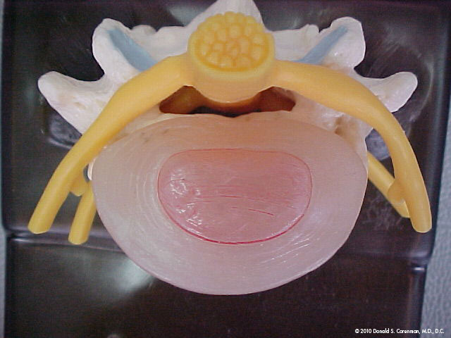
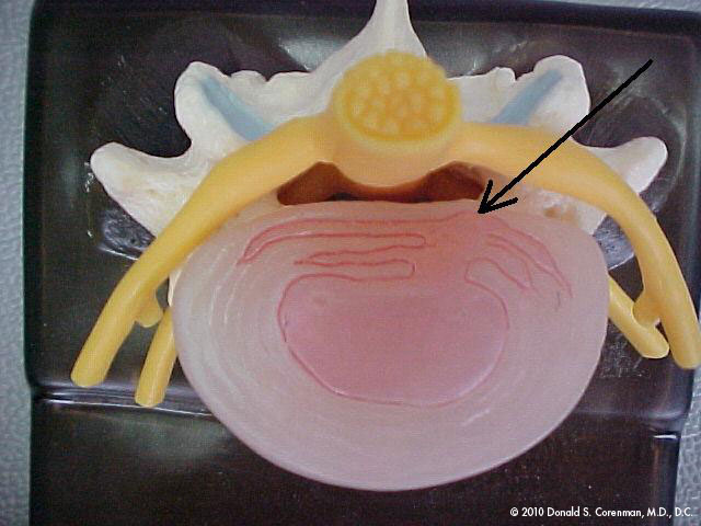
(Click to Enlarge Image) The first picture is looking down at an intact normal disc. (These pictures are of a lumbar spine disc but the cervical spine discs are almost exactly the same).The pink center is the nucleus (jelly in the donut) and the outer beige area is the annulus (donut of jelly donut fame). You can see the rings of collagen if you look carefully. Behind the disc lie the nerve roots seen in yellow. The facets in the back of the spine are seen in blue.
(Click to Enlarge Image) The second picture is of an annular tear in the back of the disc (arrow). The tear partially goes through the annulus. Note that the jelly pushes into the fibers of the annulus. The back wall of the annulus is full of pain receptors. This jelly is also neurotoxic so exposure of the pain nerves to this substance has an additional inflammation penalty.

(Click to Enlarge Image) The third pictures demonstrates a full through and through tear through the annulus and a disc herniation. The pressure of the jelly has pushed through the tear and has extruded out the back of the disc. The nerve root is compressed (arrow).
Are you suffering from symptoms of cervical degenerative disc disease?
Would you like to consult with Dr. Corenman about your condition?
You can set up a long distance consultation to discuss your
current X-rays and/or MRIs for a clinical case review.
(Please keep reading below for more information on this condition.)
Symptoms of Cervical Degenerative Disc Disease
The neck pain from the wear of these structures will typically be located in the center of the neck, the side of the neck and also may radiate into the top or middle of the shoulders. If the pain radiates down the arm or up the back of the head, this is not normally disc or facet pain but nerve pain and can occur in conjunction with neck pain. It results from compression of the nerve root which is a by-product of cervical degenerative disc disease. See the section on nerve root compression for more information on this condition.
If the disc itself loses all its jelly, the endplates start to impact into each other. This can cause the cartilage that lines the endplates to dissolve. This results in a bone on bone condition and severe neck pain can occur. This cause of neck pain is the deep dull ache that occurs after impact activities or prolonged exposure to vibration such as riding in a car or on an airplane. Intense pain can be delayed-that is, the pain can occur after the activity and patients will state that they “will pay” for the activity later.
Pain that occurs between the shoulders is normally not from the thoracic spine or shoulder but from torn disc or a slipped disc in the cervical spine.
The facets are real joints- just like the knee, hip or shoulder. Just like these joints, the facet joints can wear out and cause pain (this pain can be often related to arthritis of the neck). This type of pain is normally located in the back of the neck and aggravated by bending the head backwards but there are so many exceptions that it is difficult to make a diagnosis simply by the location of pain.
The popping, crackling, and crunching spine noises heard in the neck with motion are the irregular surfaces of the facets that rub against each other. These disturbing sounds develop in almost every person on the face of this earth. If the noises are not accompanied by pain, there is no danger. Without pain, do not pay attention to these noises. No one else can hear them but you!
Treatment of Cervical Degenerative Disc Disease
Non-Surgical
Standard treatment for cervical degenerative disc disease and cervical degenerative facet disease includes physical therapy, epidural injections and medications. Ergonomics and impact activity avoidance can be helpful. Chiropractic treatment can be very efficatious for management of these symptoms. If degenerative facets are the cause of the neck pain (about 30% of the time), dorsal rhizotomy can be effective.
Surgical
Depending upon the cause of pain, ACDF can be very effective. Normally, the disc is so degenerative that there is almost no motion of this segment. Stopping the motion with an ACDF causes almost no reduction of range of motion from pre to post-operative. Artificial discs can be used if there is still reasonable motion of the segment and there is not too much collapse of the disc. If the segment is too degenerative, artificial discs will normally not be useful here.
For more information on cervical degenerative disc disease and cervical degenerative facet disease, or for additional resources on the causes of neck pain, please contact Dr. Donald Corenman, spine specialist and neck doctor in the Vail, Aspen, Denver and Grand Junction, Colorado area.
Related Content
-
- Cervical Radiculopathy or Pinched Nerve in the Neck
- Cervical Herniated Disc
- Arthritis of the Neck and Back
- Cervical Deformity
- Cervical Degenerative Kyphosis
- Cervical Degenerative Spondylolisthesis
- Cervical Central Stenosis & Myelopathy
- Chronic Radiculopathy
- How to Describe Your History and Symptoms of Neck, Shoulder and Arm Pain
- Best Questions to Ask When Interviewing a Spine Surgeon or Neurosurgeon
- When to Have Neck Surgery