Femoroacetabular Impingement (FAI) Overview
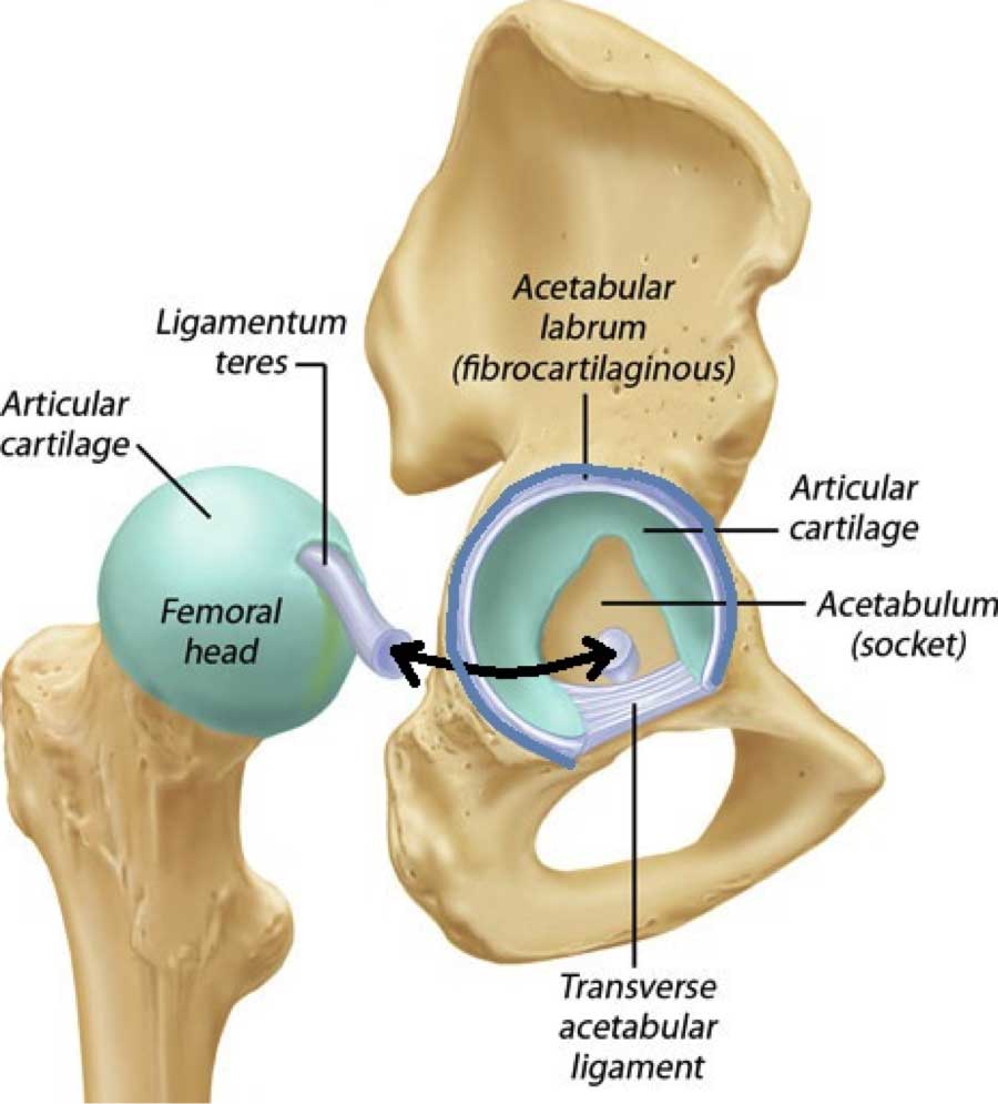
The hip joint consists of a perfectly round ball (the femoral head) that inserts into a perfectly round socket or cup (the acetabulum). Both surfaces are coated with hyaline cartilage, a firm slippery material that is bonded to both bones and reduces friction to almost nothing.
These joint surfaces are held together by a tough fibrous material called a capsule. This capsule inserts into the hip socket and contains the labrum, which is an extension of the acetabular rim (the cup). This labrum extends the depth of the hip socket and prevents the capsule from intruding into the joint.
Development of FAI
The femoral head and acetabulum are both designed to grow when we are young. This means there are growth plates present on both sides of this joint. These growth plates can displace or partially slip when developing in the growing adolescent. When the slip is substantial (called a slipped femoral capital epiphysis or SCFE) the slip becomes painful and will be addressed by a physician as the child will complain of hip pain and have great difficulty to walk.
Partial slips however may not be symptomatic but will leave the femoral head or the acetabulum “out of round”. The small “hump” remaining on the femoral head or the acetabulum due to the slip may not cause symptoms until much later in life.
When the hip is brought into certain positions, femoroacetabular impingement will occur between this boney bump and the labrum causing erosion of cartilage and a possible tearing or degeneration of the labrum.
The two different impingement types are cam and pincer mechanisms. If excess bone forms around the hip ball, it is called a cam impingement. If the impingement occurs from an overgrowth of the acetabulum (socket), it is called a pincer type.
Certain sports can induce labral degeneration. Sports that require significant hip rotation are commonly associated with labral tears. These include golf, tennis, soccer, wrestling, track events, ice hockey and ballet.
This activity causes onset of the symptoms and can lead to further erosion and eventual arthritis (wearing of the cartilage surface of the ball and socket).
(Click to Enlarge)

(Click to Enlarge) A-Normal hip (ball of femur is on the left and cup of acetabulum is on the right) B-Cam type impingement (stippled area is extra bone in the front of the ball) C-Pincer type impingement (stippled area is extra bone in the front of the socket) D-Combination of can and pincer impingement
Are you suffering from symptoms of a hip impingement syndrome?
Would you like to consult with Dr. Corenman about your condition?
You can set up a long distance consultation to discuss your
current X-rays and/or MRIs for a clinical case review.
(Please keep reading below for more information on this condition.)
Symptoms of Femoroacetabular Impingement
The majority of pain is found within the hip joint. This is the area around the front of the groin. The pain will typically be felt as a “deep” ache. Many patients will demonstrate their pain by grabbing their hip using their hand as a “c” (index finger in front and thumb in back towards the buttocks in the shape of a “c”- the C sign).
Pain can radiate to the side of the hip or even the sacroiliac joint region and can mimic lower back pain. I have seen an occasional patient presenting with pain that radiates below the knee mimicking a radiculopathy (nerve pain).
A catching or clicking sensation can occur if there is a labral tear. The catch may occur when the patient rises from a chair or exits/enters a car. Climbing or descending stairs can also cause this pain and “catching” sensation.
Sports that require frequent change of direction can cause the same pain. Getting on and off a bicycle will do this and squatting can also cause the catch or sharp momentary pain.
Eventually, prolonged sitting can cause symptoms to occur. Patients may complain of a “stiff” hip joint.
Differential Diagnosis
Where confusion can occur is with the differential diagnosis of spinal disorders that cause similar pain as femoroacetabular impingement and labral tears. That is, determining which disorder really exists so the correct treatment can be instituted.
Referral pain to the hip can occur with nerve compression of L1 through L3 and even occasionally L4. Referral pain from the L4-5 disc can cause occasional hip and groin pain.
In turn, the labral tear can refer pain to the back of the pelvis and even refer down the front of the leg below the knee (rare).
Typically, hip symptoms occur with walking, crossing over the legs, climbing and descending stairs, getting in and out of a car and on-off a bicycle. Spine symptoms occur with stretching the nerve root (from a disc herniation) or are reproduced from standing and walking. As you can see, there are overlap symptoms that are shared between hip and spine disorders.
The easiest way to differentiate between these two disorders is with an anesthetic block of the hip joint. This numbs the joint and any structures inside it temporarily. Injection of this joint is now relatively easy with ultrasound guidance and can be performed in the office. If the hip joint injection relieves symptoms for 2-3 hours, the diagnosis of hip joint disorder is normally confirmed. Hip joint injection will typically not relieve pain generated from the disc or nerve.
For additional information on femoroacetabular impingement (FAI) and its association with lower back pain, please contact the Vail, Aspen, Denver and Grand Junction, Colorado area office of back doctor and spine specialist Dr. Donald Corenman.
Related Content
- Complex Regional Pain Syndrome (CRPS) / Reflex Sympathetic Dystrophy (RSD) / Causalgia
- Dangerous Mimic of Back Pain
- Femoral Acetabular Impingement Syndrome (FAI)
- Greater Trochanteric Bursitis
- Multiple Sclerosis
- Polymyalgia Rheumatica (PMR)
- Reactive Depression
- Rheumatological Conditions That Affect the Spine
- Walking (gait) Disorders