(Click to Enlarge) 3 far lateral HNP images
Missed Diagnosis of a Far Lateral Disc Herniation Overview
This missed diagnosis of a far lateral disc herniation is unfortunately more common than would be expected. It may be that some radiologists (and some clinical physicians) are not trained to look at the lateral aspect of the MRI and identify these herniations. I see about 3-4 of these missed herniations in my office each year. The typical story is the patient with significant leg pain that shows up to an ER multiple times and are thought not to have compression of the nerve root as the MRI is read as normal. These patients might be sent away with the “there is nothing wrong with you” diagnosis.
That is the story of this patient who went to the ER multiple times. An MRI is performed and the far lateral disc herniation is not spotted. The patient was initially given pain medications and sent home because of the missed diagnosis. The pain persists as oral pain medications are not very effective on nerve compression syndromes. The patient returned to the ER multiple times due to leg pain and is then considered a “drug seeker”. This patient came to our office and was identified with a far lateral disc herniation. Subsequent surgery removed the herniation and relieved the pain.
A careful neurological examination would normally reveal a radiculopathy due to this nerve compression. That is, sensory deficit, pain with nerve stretch, reflex deficit and or motor weakness would be identified. Unfortunately, a precise examination is sometimes lost in the shuffle in the busy ER and some ER personnel do not understand a precise neurological examination. This patient had an L3 radiculopathy which manifested in thigh pain and quadriceps weakness by involving the femoral nerve, not a common presentation for an ER.
Are you suffering from symptoms of a far lateral disc herniation?
Would you like to consult with Dr. Corenman about your condition?
You can set up a long distance consultation to discuss your
current X-rays and/or MRIs for a clinical case review.
(Please keep reading below for more information on this topic.)
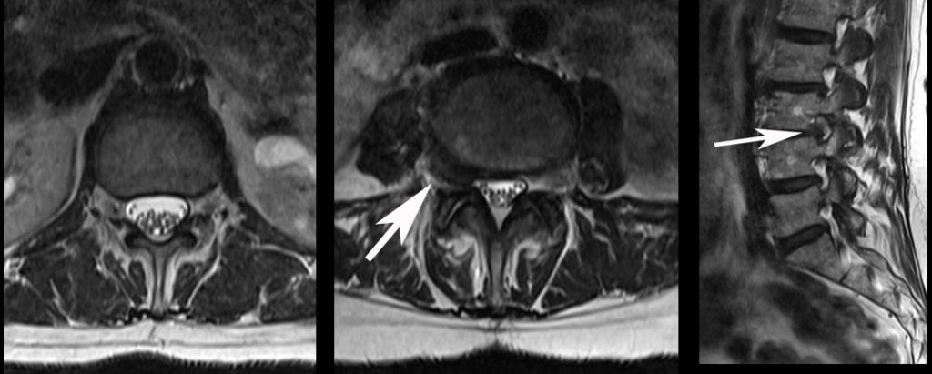
The three images are the MRI findings. The first image to the left is a “top down” view of a normal disc level above the herniation. Compare to the second image with the white arrow. This arrow points to the black mass that is compressing the nerve root. The final image is the “side view” of the spine with the arrow pointing to the black mass in the foramen at L3-4. Compare to the foramen above and below this level. The nerve is compressed as seen by the gray structure above the black mass.
For additional resources on a missed diagnosis of a far lateral disc herniation, please contact the office of Dr. Donald Corenman, back doctor and spine specialist offering diagnostic and surgical second opinions to patients in the USA and around the world.