What is the Atlanto-Occipital Joint?
The atlanto-occipital joint (O-C1) is formed by the occipital condyles and the upper articular facets of C1. These paired joints each look like a saddle and both are stabilized by multiple ligaments and the articular capsules of O-C1.
The O-C1 joint is responsible for the most flexion/extension of any vertebral segment with 25 degrees of motion. There is a small amount of rotation (5 degrees) that also occurs here.
The basion is the front edge of the foramen magnum and the opisthion is the rear edge, important bony contact points for the stabilizing ligaments of this joint.
The stability of the atlanto-occipital joint stems from multiple structures. The aforementioned joint capsules of the O-C1 joint create substantial stability. The other ligaments are noted here from front to back.
How the Atlanto-Occipital Helps with Neck Stabilization
The anterior atlanto-occipital membrane (a continuation of the anterior longitudinal ligament) attaches from the anterior arch of the atlas to the anterior aspect of the basion (the front of the foramen magnum). This ligament works to prevent excessive neck extension.
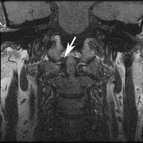
The twin alar ligaments attach from the lateral aspect of the odontoid process (dens of C2) to the medial occipital condyles on either side. These ligaments limit flexion and rotation at the atlanto-occipital joint.
The apical ligament attaches from the tip of the odontoid process (C2) to the basion. It is sandwiched between the alar ligaments and the cruciate ligament complex. This ligament is not significant and does not contribute to the stability of O-C1.
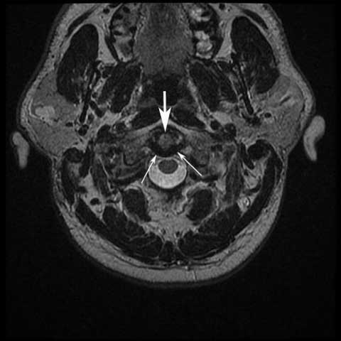
The cruciate ligament complex is just posterior to the odontoid process originating from the back of the dens. The whole ligament complex looks like a cross (cruciate). The superior band stabilizes the odontoid to the basion. The transverse band (better know as the transverse ligament) is the strongest portion of the cruciate ligament complex and loops around the odontoid, attaching to the lateral masses of the atlas. This ligament is the most important stabilizer of C1-2. The inferior band is a continuation of the superior band and connects to the body of C2.
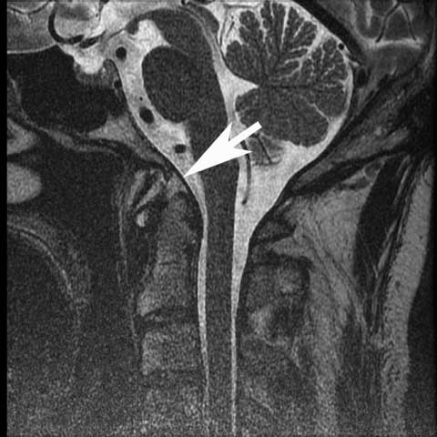
The tectorial membrane is an extension of the posterior longitudinal ligament and lies posterior to the cruciate ligament complex. This ligament attaches from the dens to the anterior foramen magnum. Due to its position, this ligament limits both excessive flexion and extension.
Jumping to the back of the spinal canal, the posterior atlanto-occipital membrane attaches from the occipital bone to the posterior arch of the atlas. It is a continuation of the ligamentum flavum and a homolog of the anterior occipital membrane.
The ligamentum nuchae is a continuation of the supraspinous ligament and spans from the posterior occiput to the spinous process of C1. This ligament serves to limit excessive neck flexion.
Related Links
- Normal Spinal Alignment
- Anatomy of Thoracic Spine
- Anatomy of Lumbar Spine
- When to Have Lower Back Surgery
- When to Have Neck Surgery
- How to Describe Your History and Symptoms of Neck, Shoulder and Arm Pain
- How to Describe Your History and Symptoms of Lower Back and Leg Pain
- Anatomy and Motion of the Cervical Spine
(Click to Enlarge) Alar ligaments attach from the odontoid to the condyles of the skull. Large arrow odontoid, small arrows alar ligaments.
(Click to Enlarge) Tectorial membrane arrow points to membrane continulation of PLL.
(Click to Enlarge) Transverse ligament top down of odontoid. Large arrow and ligament small arrows.


