The thoracic spine is the area between the neck and lower back that supports the chest. The shape of the thoracic vertebrae is similar to those in the anatomy of the lumbar spine but reduced in size. Again, just like its neighbors above and below, the disc in front separates the two vertebral bodies and the facets in back guide the motion of the spine. The discs in the thoracic spine have the same anatomy as the lumbar spine but are thinner and somewhat inflexible. The major difference between this area and the lumbar spine is that ribs attach to each thoracic vertebra and this makes this area of the spine very stiff.
The alignment of the spine differs from the neck (the cervical spine) and lower back (the lumbar spine). From front to back, the spine should be straight (a curve greater than 10 degrees would be considered a scoliosis). From the side view, the spine should have a front to back curve called a kyphosis. This curve ranges from 20-40 degrees and is important to balance out the entire spine. This curve along with the opposite curves in the neck and lower back balance the head over the pelvis.
The central canal contains the spinal cord. Compression of the spinal cord will cause symptoms similar to cervical stenosis with myelopathy (see that section for description) but the arms will not be affected. Unlike the cervical and lumbar spines, the nerves of the thoracic spine do not exit into the arms or legs, but do exit into the chest wall.
The discs all lose their blood supply by the age of 8, so just like all discs, injuries to the discs are permanent and cumulative. Because of the rib attachments, discal tears are uncommon and herniations are rare. An MRI can help a spine specialist look at the thoracic spine.

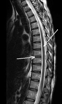
(Click to Enlarge Image) This is a lateral (sagittal) MRI of a normal thoracic spine. The upper white arrow points to the spinal cord and the lower white arrow points to the normal disc space. You can see that there is no compression of the spinal cord and all the discs look the same (no degenerative changes). The spinal cord fades out of the image as this individual does not have a perfectly straight spine (normal).
