An Overview of
The interesting crystalline material structure of bone is the underpinning for the development of stress fractures. Fatigue fractures of a long bone are not uncommon under significant repeated load. It makes us realize that long bones are just like any brittle structure that can be overstressed. This goes back to the stress/strain curves in materials management in physics. This condition even occurs in the spine (see pars fractures and isthmic spondylolisthesis).
Bone, like any crystalline structure, has an absolute fatigue point for gross failure (a fall from a height breaking the leg). There is however the possibility of cumulative stress from constant impact that does not break the bone fully. Tiny fractures in the substance of the bone can occur that do not individually reach the gross failure point.
These stresses accumulate over time with repeated loads. This is a time and stress related function. Stress fractures overwhelm the ability of the bone to heal without rest (time).
An analogy would be taking a wire coat hanger and bending it continuously until it breaks. No one individual bend causes failure but the accumulation of bends breaks the wire. It is remarkable that bone has a self-healing process and can recover from these less than full structural failure injuries given time and rest.
Groups at Risk for
Two groups that have the greatest risk of are the athletic and the military populations. In either population, prolonged impact activities (running, weight lifting and calisthenics) set the stage for these overuse fractures.
Insufficiency fractures due to inferior bone stock generally found in the elderly due to osteoporosis are also at high risk. These fractures also occur in the thoracic and lumbar vertebra.
Location of Lower Extremity Fractures
The typical location of these lower extremity fractures is generally found in long tubular bones. Tibial stress fractures, the most common are at 50% of all stress fractures. Metatarsal stress fractures (foot) occur under 20% of the time and femoral shaft and neck fractures exist at about a 10% occurrence rate.
There are potential risks and different healing outcomes for fractures in different locations. As would be expected, fractures with reduced vascularity due to location and anatomy (the anterior tibia and navicular bone in the foot) had poorer outcomes due to this relative lack of blood supply. If a fracture of the femoral neck is allowed to develop and then displace, the chance of avascular necrosis (bone death) to the femoral head is very high due to the retrograde vascularity of the femoral head.
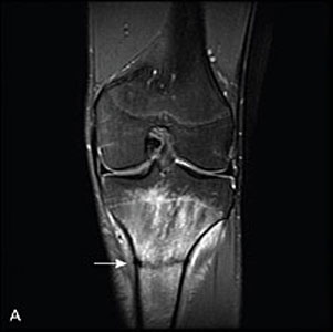
(Click to Enlarge Figure A)

(Click to Enlarge Figure B) Figure A & B – Note the white signal in the tibia (lower bone in the picture) and compare this with the black signal in the higher up femur. The white signal is fluid within the bone due to the small stress fractures of the small inside bones (trabecula). The arrow points to a full black fracture line in the tibia that occurred after enough stress fractures accumulated to allow this obvious full fracture to propagate.
Are you suffering from symptoms of a lower extremity fracture?
Would you like to consult with Dr. Corenman about your condition?
You can set up a long distance consultation to discuss your
current X-rays and/or MRIs for a clinical case review.
(Please keep reading below for more information on this condition.)
Symptoms of
Symptoms of stress fractures of the lower extremities include localized pain with impact activities that increase in intensity and with prolonged impact or stress exposure. If the fracture develops further, localized swelling and tenderness are noted. Day to day pain with normal activities is an indicator of a progressive fracture.
Risk factors are identified with a thorough history and physical examination. These factors include female status, menstrual irregularities and less muscle mass in the lower extremities. In fact, females who had five or less menses per year had almost double the risk of stress fractures compared to those who had a normal menstrual cycle. Interestingly, oral contraceptives reduced the chances of developing stress fractures.
Diagnosis of
This potential diagnosis has to be on the practitioner’s radar screen to be able to make the diagnosis and prevent serious sequelae. Complications of a missed diagnosis include a complete fracture of the tibia or even the need for a hip replacement due to avascular necrosis of the femoral head.
Uncommon questions such as how many menses the female has had in the last year are important to know. Practitioners need to understand the significance of this question (among others) with a young athletic female who complains of leg pain. Sir William Osler’s famous quote, “You find what you know and you see what you look for” applies here (as well as in most circumstances in medicine).
Other than the history, there are few physical examination findings that can lead to a diagnosis. Local tenderness can be a clue to the presence of a lower extremity fracture. If there is swelling at the site, this indicates a more substantial and progressive stress fracture.
The MRI is the leading tool for confirming diagnosis after a suspicion of stress fracture is considered. Prior to the development of MRI technology, bone scan and CT scan were utilized but had a much lower sensitivity for this injury. Now, with the ability to visualize the interstitial fluid within the bony matrix from the fractures of the trabecula and laminar layers of the cortical bone, identification can occur much earlier. This is especially noted with STIR images.
Clinical Relevance
Unexplained lower extremity localized pain in athletic or military personnel should be potentially considered a stress fracture with the resultant work-up necessary to diagnose this disorder. MRI of the painful region is required to determine the presence of a stress fracture.
Lower extremity insufficiency fractures due to osteoporosis are more common than we estimate. The alert practitioner needs to be especially careful to identify and treat these patients. A positive log roll test of the hip may be an early warning sign of a femoral neck stress fracture. Any unexplained lower extremity pain that increases with loading has to be considered a possible fatigue fracture in an elderly patient.
For additional information on
Related Content
- When to Have Lower Back Surgery
- Causes of Lower Back Pain
- Normal Spinal Alignment
- Lumbar Spine Anatomy
- How to Describe Your History and Symptoms of Lower Back and Leg Pain
- Best Questions to Ask When Interviewing a Spine Surgeon or Neurosurgeon
- Degenerative Spondylolisthesis
- Isolated Disc Resorption-Lumbar Spine (IDR)
- Lumbar Spinal Stenosis (Central Stenosis)