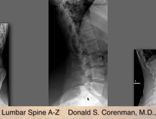
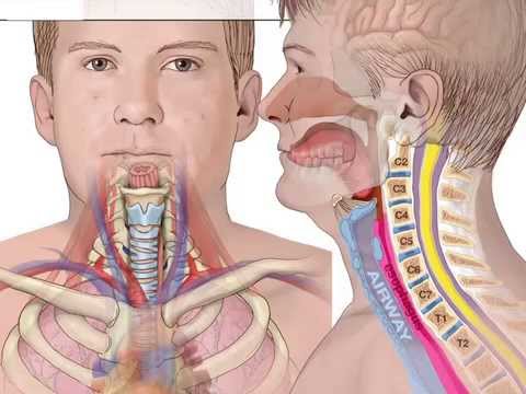
This video–Understanding X-rays of the Cervical Spine–is designed for the practitioner who has access to x-rays for diagnosis of neck pain and neck disorders (See the anatomy section for the anatomy of the cervical spine). Cervical X-rays can deliver so much information in so few films. One can look at the health of the discs, uncovertebral joints, facets, nerve root foramen, alignment, spinal canal diameter, and even the occiput-C2 joint complex. This test is the only real way to determine how gravity affects the spine (MRIs and CT scans are performed lying down and the stand-up MRIs are of poor quality). Flexion and extension views add to the knowledge of motion of the spine and oblique films can identify bone spurs in the foramen.
Share This Story, Choose Your Platform!
About the Author: Donald Corenman, MD, DC
Donald Corenman, MD, DC is a highly-regarded spine surgeon, considered an expert in the area of neck and back pain. Trained as both a Medical Doctor and Doctor of Chiropractic, Dr. Corenman earned academic appointments as Clinical Assistant Professor and Assistant Professor of Orthopaedic Surgery at the University of Colorado Health Sciences Center, and his research on spine surgery and rehabilitation has resulted in the publication of multiple peer-reviewed articles and two books.
