Anterior Cervical Decompression and Fusion (ACDF) Overview
The anterior cervical decompression and fusion (ACDF) procedure is performed for cervical instability, painful degenerative disc disease, bone spur compressing the nerve root or spinal cord, herniated disc or even fracture.
The purpose of this surgery is to restore the collapsed disc to the original height before degenerative changes occurred, remove herniation or bone spur to free the nerves or spinal cord and stabilize the segment to prevent further detrimental motion. This surgery is performed on the cervical spine and is performed with anterior surgery (from the front of the neck).
A small incision is made in a Langer line-a small wrinkle in the skin that runs transversely in most individual’s necks. There is a very thin vestigial muscle called the platysma (one that might have served a purpose in prehistoric times and is important in birds but does not help humans now) that is incised (and repaired at the end of the case). After that, there are no structures in the way and the surgeon can literally “fall” down an interval directly onto the front of the spine of the neck. The trachea and esophagus are retracted medially and the carotid sheath is retracted laterally.
The disc is identified by x-ray, two posts are placed into the vertebra above and below the disc and the vertebra are distracted apart. The disc quite commonly will have a through and through tear in it (the annulus). The front of the annulus is removed and whatever debris is left in the disc space is removed. There is normally a thin layer of cartilage on the surfaces of the vertebra and this is removed. A burr (similar to a $65,000.00 dremel) is used to fashion the endplates parallel and thin down the lips at the back side of the vertebral bodies (the posterior and lateral uncovertebral joints for those of you on Jeopardy).
Are you a candidate for anterior cervical decompression and fusion (ACDF)?
Would you like to consult with Dr. Corenman about your condition?
You can set up a long distance consultation to discuss your
current X-rays and/or MRIs for a clinical case review.
(Please keep reading below for more information on this treatment.)
Using very small tools, the spurs are removed and the herniations are removed. A graft is then used to be placed in between the vertebral bodies to cause them to fuse together. The graft can be from your own bone (ICBG-iliac crest bone graft) or a PEEK cage which acts as a spacer and is filled with your own bone and demineralized bone matrix (DBM). The PEEK cage is imbedded with a thin film of titanium (see image) which is biologically active and allows bone to adhere to it. The discussion as to which graft to be used will be made before the anterior cervical decompression and fusion (ACDF) surgery is scheduled. Both grafts have advantages and disadvantages that will be discussed.
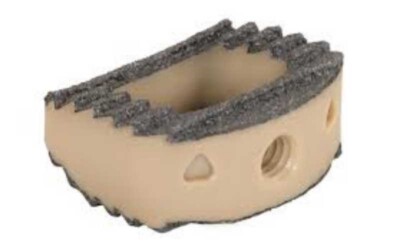
PEEK Cage
A small titanium plate is placed on the two vertebra, the incision is sutured, the patient is placed in a neck collar and sent to the recovery room.
For additional resources on anterior cervical decompression and fusion (ACDF), or for information on other orthopedic spine treatment options, please contact the office of Dr. Donald Corenman, neck doctor and spine surgeon offering diagnostic and surgical second opinions to patients in the USA and around the world.
Related Content
- Anterior Cervical Decompression & Fusion (ACDF)
- Artificial Disc Replacement (ADR) for Cervical Spine
- Cervical Laminectomy, Laminoplasty and Posterior Cervical Fusion
- Posterior Cervical Foramenotomy
- Myths of Laser Spine Surgery
- How to Describe Your History and Symptoms of Neck, Shoulder and Arm Pain
- Best Questions to Ask When Interviewing a Spine Surgeon or Neurosurgeon
- When to Have Neck Surgery
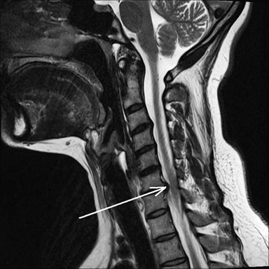
(Click to Enlarge Image) Lateral cervical MRI of the disc herniation noted by the white arrow.
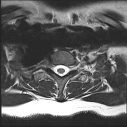
(Click to Enlarge Image) Axial view (bottom up) of a normal level without disc herniation.
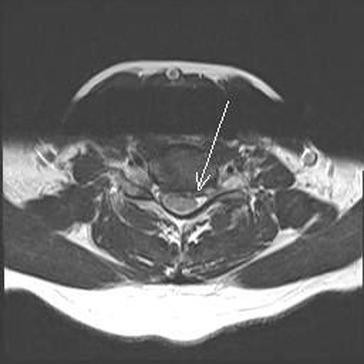
(Click to Enlarge Image) Axial view of the disc herniation level (white arrow).

(Click to Enlarge Image) Lateral X-ray of the post-operative ACDF.