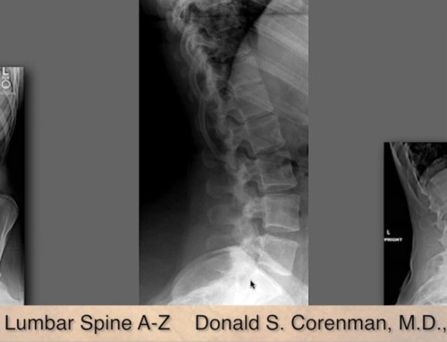
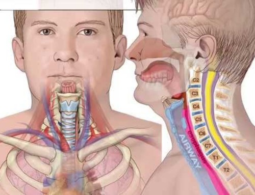
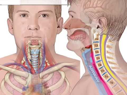
This video–Understanding an MRI of Cervical Nerve Compression–relates to the cervical spine (neck) and is designed for the primary care physician or specialist such as a Chiropractor or Physical Therapist to use to learn how to read and gain an understanding of an MRI of the cervical spine with nerve root compression. These foramen (or holes the nerves exit from) take some time to visualize and understand what is normal and what is narrowed or compressed. The uncovertebral joint can enlarge by bone spur formation and can compress the nerve. A disc herniation can also compress the nerve and that has a different appearance from the uncovertebral joint spur. Please see the video Cervical Foraminal Stenosis for an explanation of the spur and what it looks like in an anatomic model. Of course, the physical examination and patient complaints are extremely important as just because there is a compressed nerve does not mean there will be symptoms.
Share This Story, Choose Your Platform!
About the Author: Donald Corenman, MD, DC
Donald Corenman, MD, DC is a highly-regarded spine surgeon, considered an expert in the area of neck and back pain. Trained as both a Medical Doctor and Doctor of Chiropractic, Dr. Corenman earned academic appointments as Clinical Assistant Professor and Assistant Professor of Orthopaedic Surgery at the University of Colorado Health Sciences Center, and his research on spine surgery and rehabilitation has resulted in the publication of multiple peer-reviewed articles and two books.
